Ankle injuries are common, and among them, malleolar fractures are particularly significant due to their impact on mobility and daily activities. Malleolar fractures involve the bony prominences of the ankle, known as the malleoli, and can range from minor cracks to severe breaks. At Prisk Orthopaedics and Wellness, PC, we aim to provide comprehensive care for these injuries, ensuring optimal recovery and return to normal function.
Recognizing a Malleolar Fracture
Knowing when your ankle is broken is crucial for seeking timely medical intervention. Here are some signs and symptoms that might indicate a malleolar fracture:
- Severe Pain: Sudden, intense pain at the time of injury is a primary indicator. The pain may worsen with movement or when trying to bear weight.
- Swelling and Bruising: Rapid swelling and bruising around the ankle are common signs of a fracture.
- Deformity: A visible deformity, such as an abnormal angle or misalignment of the ankle, strongly suggests a fracture.
- Inability to Bear Weight: If putting weight on the injured ankle is too painful or impossible, it may be fractured.
- Tenderness: Tenderness around the bony parts of the ankle, especially the malleoli, can indicate a fracture.
If you experience any of these symptoms following an ankle injury, it’s crucial to seek medical attention promptly. Delaying treatment can lead to complications and prolonged recovery.
Advanced Diagnostic Tools at Prisk Orthopaedics and Wellness, PC
Accurate diagnosis of malleolar fractures is essential for effective treatment and recovery. At Prisk Orthopaedics and Wellness, PC, we utilize state-of-the-art diagnostic tools to ensure precise identification of fractures, including those that are often missed by standard imaging techniques.
LineUP CT Scan by Curvebeam
Our LineUP CT scan by Curvebeam offers advanced imaging capabilities that allow us to diagnose occult fractures that many clinicians may not detect. This high-resolution imaging technology provides detailed views of the bone structures, enabling us to identify even the smallest fractures and ensure comprehensive treatment planning.
Weaknesses in Existing Clinical Literature
The available clinical literature on BPC-157 is sparse and fraught with methodological weaknesses. Most of the studies that are often cited to support the use of BPC-157 are preclinical, conducted on animal models. While these studies suggest potential benefits, they do not provide the level of evidence needed to confirm safety and efficacy in humans.
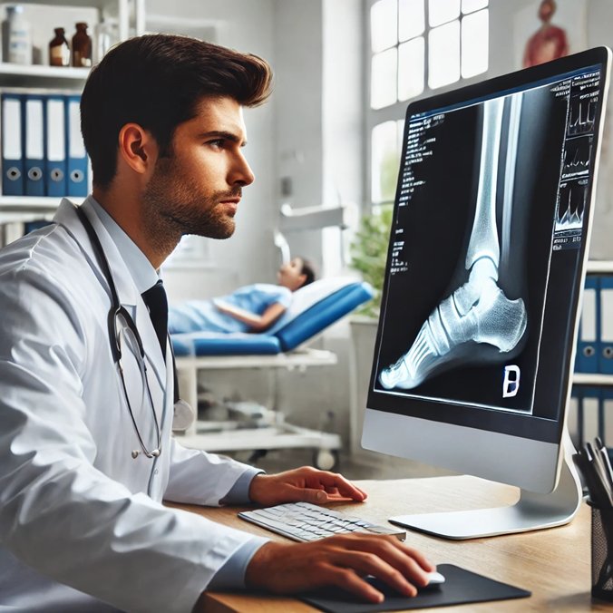
Cesium Plate Digital X-Ray
We also boast cesium plate digital X-ray technology, providing the highest quality X-rays in the Pittsburgh region. This cutting-edge imaging system delivers superior image clarity, allowing for more accurate assessments of bone injuries. The enhanced resolution helps in detecting subtle fractures and aids in precise treatment decisions.
Treatment Options for Malleolar Fractures
Treatment for malleolar fractures varies depending on the severity and type of fracture. At Prisk Orthopaedics and Wellness, PC, we offer both nonoperative and operative treatments to cater to individual patient needs.
Nonoperative Treatment
Some malleolar fractures can be managed without surgery, particularly if the bones are not displaced. Nonoperative treatment typically includes:
- Immobilization: Using a cast or a boot to immobilize the ankle, allowing the bones to heal naturally. This prevents movement and ensures proper alignment during the healing process.
- R.I.C.E. Protocol: Rest, Ice, Compression, and Elevation are essential components of initial care. Elevation helps reduce swelling by promoting fluid drainage, while ice and compression minimize swelling and pain.
- Weightbearing Restrictions: Initially, you may be advised to avoid putting weight on the injured ankle. Gradually, as healing progresses, partial weightbearing with crutches or a walker may be introduced under medical supervision.
Operative Treatment
In cases where the fracture is displaced or unstable, surgical intervention may be necessary. Orthopedic implants, such as plates and screws, are used to realign and stabilize the bones. The surgical process typically involves:
- Open Reduction: The surgeon makes an incision to access the fractured bones, realigning them to their proper position.
- Internal Fixation: Plates, screws, or other implants are used to secure the bones in place, ensuring stability and proper healing.
The Importance of Perioperative Care
Whether treated nonoperatively or operatively, perioperative care is crucial for successful recovery. Here’s a detailed look at the essential aspects of care during this period:
Elevation
Keeping the injured ankle elevated, especially in the initial stages, is vital for reducing swelling and pain. Elevation helps drain excess fluid from the injury site, minimizing inflammation and promoting faster healing. Aim to keep the ankle elevated above heart level as much as possible, particularly in the first 48 hours post-injury or surgery.
Following Weightbearing Restrictions
Adhering to weightbearing restrictions is critical to prevent further damage and ensure proper healing. Your orthopedic surgeon will provide specific guidelines on when and how to bear weight on the injured ankle. Ignoring these restrictions can lead to complications such as delayed healing, malalignment, or re-injury.
Physical Therapy
Engaging in a structured physical therapy program is essential for regaining strength, flexibility, and function in the ankle. Physical therapy helps:
- Restore Range of Motion: Gentle exercises and stretches to improve mobility.
- Strengthen Muscles: Strengthening the muscles around the ankle to support the joint.
- Improve Balance: Exercises to enhance balance and prevent future injuries.
Pain Management
Managing pain effectively is crucial for comfort and mobility during the healing process. Your physician may prescribe pain medications or recommend over-the-counter options. Additionally, ice therapy and elevation can help reduce pain naturally.
Monitoring for Complications
Regular follow-up appointments are necessary to monitor the healing process and detect any potential complications early. X-rays may be taken to ensure the bones are healing correctly and that the implants (if used) remain in place. Be vigilant for signs of complications, such as increased pain, swelling, redness, or fever, and report these to your physician immediately.
Conclusion
Malleolar fractures can significantly impact your mobility and quality of life. However, with proper diagnosis, appropriate treatment, and diligent perioperative care, you can achieve a successful recovery. At Prisk Orthopaedics and Wellness, PC, we are committed to providing personalized care to ensure the best outcomes for our patients. Our advanced diagnostic tools, including the LineUP CT scan by Curvebeam and cesium plate digital X-ray, allow us to detect and treat even the most subtle fractures effectively. If you suspect a malleolar fracture or need expert care for an ankle injury, contact us for a comprehensive evaluation and treatment plan. Your road to recovery starts here.